I thought that this would be helpful to those that are looking for answers or those that would like to see what radiographic evidence looks like and how it is defined...
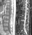
(a) T1-weighted and (b) short tau inversion recovery (STIR) magnetic resonance images of lumbar and lower thoracic spine in psoriatic arthritis. Signs of active inflammation are seen at several levels (arrows). In particular, anterior spondylitis is seen at level L1/L2 and an inflammatory Andersson lesion at the upper vertebral endplate of L3. The fourth, fifth and sixth vertebra contains certain peculiarities, which are detailed below.
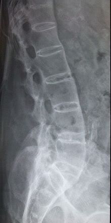
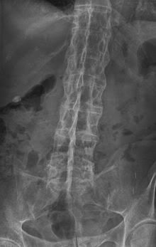
Radiology shows the appearance of "fluffy, new" bone or bridging/fusion.